Three Part Article: Ear Disease in Dogs
by Nancy Kay, DVM | Three Part Article taking from her popular series Speaking for Spot
EAR DISEASE IN DOGS - PART ONE | Anatomy Of The Ear Canal
EAR DISEASE IN DOGS - PART TWO | Common Ear Problems
EAR DISEASE IN DOGS - PART THREE | Dealing With Repeat Symptoms
EAR DISEASE IN DOGS - PART ONE
 If you are a dog lover, chances are one of your canine companions has experienced an ear infection. No fun, right? And if your dog has suffered with recurrent ear infections, chances are you’ve felt like pulling your own hair out!
The following information is intended to enhance your understanding of the canine ear and help you get the help you need if your dog develops ear issues.
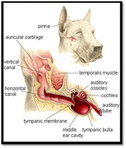
The anatomy of the canine ear canal
I invite you to join me on an imaginary trip. Pretend you are miniaturized to the size of Tom Thumb. Now slip beneath your dog’s earflap and sit yourself down on the ledge of the opening to the ear canal. Give yourself a little push-off and then let gravity launch you down what feels like a giant slip and slide. Here is what you will see along the way.
The first part of your voyage is within the vertical ear canal, aptly named because of its rather steep descent. You are sliding down a wide-open tunnel with smooth pink surfaces. You come to an abrupt halt as you arrive at a rather sharp bend within the tunnel. You stand and take a few short steps that bring you to another descent, this one more gradual. You are now within the horizontal portion of the external ear canal. Looking forward you see a glistening, semi-transparent membrane that fills the entirety of the tunnel ahead. Aha! This must be the eardrum (tympanic membrane). It appears quite thin so you muster up some speed and run straight towards it. Your force and momentum cause the eardrum to rupture and you topple forward into a large cavernous space.
The middle ear
You’ve now entered the tympanic bulla (middle ear cavity), an open and empty, smooth surfaced cave created out of bone. It’s not easy, but you manage to climb out of this cavern, and as you approach the top you notice multiple small bones (ossicles that are responsible for transmitting sound) along with what looks like a thin window shade called the cochlear window. You poke your head through the shade and find yourself peering into yet another space. Bold little traveler that you are, you climb on in.
The inner ear
You have entered the inner ear a rather small and crowded space filled with some really crazy looking labyrinthine structures. Some of them are responsible for transmission of sound to the brain, others for maintenance of balance. You see a white ropy structure that is a nerve leading directly into the brain. Now wait just a minute before you grab hold of that nerve! I think you’ve done enough for one day!
What you’ve observed
Exhausted as you are after your incredible journey you’ve likely gained some new knowledge about the canine ear:
EAR DISEASE IN DOGS - PART TWO
 If your dog suffers from ear disease, this blog’s for you! Ear problems can be so darned frustrating to deal with, primarily because they are so prone to recurrence. Part One of this series focused on the anatomy of the canine ear canal. If you haven’t had a chance to read this I encourage you to do so. Observing the length and slope of the external ear canal will help you understand why dogs are prone to otitis externa (inflammation in the external ear canal) and why it can be difficult to treat.
Why is it that some dogs go through an entire lifetime without a single ear problem, yet others become lifelong repeat offenders? Here are some predisposing factors:
Allergies
Allergies are commonplace in dogs. Some develop allergies to food ingredients, others to environment allergens such as dust, pollens, and molds. While most allergic dogs have itchy skin, some experience inflammation within the ear canals as their only symptom. This inflammation causes production of excess cerumen (ear wax) which happens to be an ideal culture media for the growth of yeast and bacterial organisms.
Identifying and appropriately treating the underlying allergies are necessary to eradicate the chronic ear problems they cause. Doing so may involve a hypoallergenic food trial (strict adherence to a novel protein diet for six to eight weeks) or specific testing to identify which environmental allergies are at play (skin testing preferred to blood testing).
Underlying diseases that affect the skin
The lining of the ear canals is truly an extension of the skin, so it makes sense that diseases that cause skin inflammation may have the same effect on the ear canals. As discussed above, allergies are a classic example. Other diseases that can affect both skin and ears include seborrhea, autoimmune diseases, mites, and hormonal imbalances such as diabetes, hypothyroidism (inadequate production of thyroid hormone), and Cushing’s disease (overproduction of adrenal gland hormones). Treatment of the underlying primary disease is the best bet for resolving the ear problems.
Moisture within the ear canal
Small numbers of yeast and bacterial organisms reside within the normal ear canal. Add moisture to the mix and the populations of these microorganisms can multiply resulting in infection.
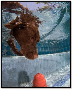
When water enters the ear canal it tends to stay put, thanks to gravity working in conjunction with the length and slope of the ear canal. No matter how much head shaking occurs or how many cotton balls are used to soak up the surface water, that ear canal is going to stay wet following swimming and bathing.
The options for dealing with this situation are to prevent the ear canals from ever getting wet (you try suggesting this to someone with a Labrador and a backyard swimming pool), or the consistent application of “drying agent” into the ear canals after they get wet. Ask your veterinarian for a product recommendation. The recipe for a homemade drying agent consists of one part white vinegar, one part water, and one part 70% isopropyl alcohol (avoid the 90% variety). Please do notuse this concoction in your dog’s ears before discussing it with your veterinarian.
Gently place a wad of cotton balls within the opening to the external ear canals prior to bath time. Once they are place coat the outer surface of the cotton balls with some petroleum jelly to help repel water. Be sure to remember to remove them when bath time is over!
A growth or foreign body within the ear canal
Any time normal anatomy is disrupted by something that shouldn’t be there, infection is likely to result. The ear canal commonly responds to the presence of a mass or foreign body in this fashion. This is one of the reasons it is so important for a veterinarian to visually inspect the entirety of an infected ear canal using an instrument called an otoscope. Removal of the mass or foreign body is the key to treating the secondary ear infection.
Narrowed (stenotic) ear canals
The normal ear canal is a wide-open structure. When narrowed, it prevents normal air circulation and predisposes to accumulation of waxy discharge. Both of these factors create the perfect storm for infection to occur. Some dogs are born with stenotic ear canals. For others narrowing is a sequela to chronic inflammation that causes thickening of the tissues lining the ear canal. In severe cases, surgical revision to “open up” the ear canal may be necessary.
Are you wondering why I did not add “hairy ear canals” to this list of factors predisposing to canine ear disease? In the good ole’ days we used to torment dogs by stripping the hair out of their ear canals thinking this would prevent infection. Now we know that doing so actually creates inflammation that can then lead to infection. With rare exception, hair removal from the ear canals is a big “no-no”.
EAR DISEASE IN DOGS - PART THREE
 Veterinarians in general practice are just about guaranteed to see at least a few dogs every week suffering from ear problems. Why is this so common? Multiple predisposing factors and theanatomy of the canine ear canal create the “perfect storm” for inflammation and infection to occur.
Although several different diseases can affect the external ear canal, they all tend to produce pretty much the same symptoms: head shaking and ear scratching +/- redness and discharge within the ear canal. Given their generic nature, one cannot rely on a dog’s symptoms to disclose the underlying cause of the problem.
Honing in on the diagnosis typically requires two steps, both performed by a veterinarian. First, the entire length of the external ear canal is visualized using an instrument called an otoscope. This rules out the presence of a mass or foreign body and confirms that the eardrum has not been ruptured. A torn eardrum will require alteration of normal ear cleaning procedures and the types of medications used. A successful otoscopic exam can be a difficult if not impossible task on a dog with a painful ear filled with discharge. Sedation or even anesthesia may be required.
The second diagnostic step is examination of discharge from the ear canal under the microscope, looking for bacteria, yeast, or mites. If allergies are at play, inflammatory cells may be the only finding. The most appropriate medication is chosen on the basis of these findings.
This article will discuss the different types of ear canal disease dogs develop. The next article in this series will provide pointers for dealing with dogs who tend to be repeat offenders.
Allergic ear disease
Any dog can suffer from allergies, but some breeds are particularly predisposed. Terriers of all types are notorious allergy sufferers along with Dalmatians, Lhasa Apsos, Shar Peis, Bulldogs, and Labrador Retrievers. Unlike humans who develop stuffy noses, dogs with allergies tend to have itchy skin and/or inflammation within their ear canals. This ear inflammation invariably leads to secondary bacterial or yeast infections. In fact, for some allergic dogs recurrent ear infections may be their one and only symptom.
Dogs can develop allergies to things in their environment (the dog’s version of hay fever) or to ingredients in their food. Anti-inflammatory medications can be used in the ear, but true resolution of this ear problem relies on successful treatment of the underlying allergy. This may involve dietary trials, skin testing to determine responses to environmental allergens, and/or treatment with medication to control the allergic response. Other medication will be needed if the allergy has given rise to a secondary bacterial or yeast infection (see below).
Bacterial infections
Bacteria are normal residents on the skin surface as well as within the external ear canals. It is when these “normal bacteria” overpopulate or different types of bacteria set up housekeeping that an ear infection arises.
Bacterial infections are most commonly treated with topical antibiotics (placed directly within the ear canal). Rarely are oral antibiotics necessary. Initial flushing (cleaning) of the ear canal may be performed to remove as much discharge from the ear canal as possible, but only if it is known with certainty that the eardrum is in tact (ear flushing should not be done at home without approval and instruction from a veterinarian).
A culture to identify the type of bacteria present along with antibiotic sensitivity testing may be warranted for dogs with recurrent infections or when there is a lack of response to the antibiotic prescribed. Bacterial ear infections are rarely contagious from dog to dog.
Yeast infections
Malassezia pachydermatis is the technical name for the yeast that normally lives within a dog’s ear canal. Symptoms of infection occur when these microorganisms proliferate in response to underlying factors such as allergies or moisture.
Yeast infections are treated with initial ear cleaning (if the eardrum is in tact) and medicated eardrops or ointment to reduce the yeast population back down to their normal numbers. Sometimes, an oral antifungal medication is prescribed. Yeast infections are not contagious from dog to dog.
Ear mites
Whereas mites are one of the most common causes of ear disease in cats, they are a relatively uncommon cause in dogs. Mange mites (demodex and sarcoptes) cause skin disease that can involve the ears. The true “ear mite” that affects only the ears is Otodectes cyanotis. These microscopic critters are highly contagious between dogs and cats. They march around within the ear canal biting, laying eggs, and generally wreaking havoc. A cure is accomplished by removal of excess debris from the ear canal in conjunction with a topical mite killing medication and/or use of a systemic anti-parasite product. All dogs and cats within the household should be treated simultaneously.
Foreign bodies
The most common foreign body that makes its way into a dog’s ear canal is a plant awn referred to as a foxtail. If you live west of the Mississippi, your dog may be exposed to them during the late spring and summer months. Once in the ear canal, the foxtail’s barbs along with the slope of the ear canal prevent the darned thing from being dislodged, no matter how much head shaking the poor dog does.
Ticks sometimes migrate down into the ear canal where they become a “foreign body” until mealtime is over and they climb back out. Other foreign objects I have removed from doggie ears include nonfoxtail types of plant material, dirt, sand, hair, and an occasional “something or other” dropped in the ear canal by a curious child.
Keep in mind that gravity will carry the vast majority of foreign bodies down into the ear canal beyond view from the surface. Their removal is best accomplished by a veterinarian. An otoscope is used for visualization and a snare or an instrument called an “alligator forceps” is used to remove the foreign body. Even the best-behaved dogs may require sedation or anesthesia for this procedure. On occasion, a foreign body manages to perforate the eardrum. If a foreign body is not removed it almost invariably incites a secondary infection.
Masses
Masses and polyps occasionally arise within the ear canal. It is usually the secondary infection they cause that first attracts attention. Ear canal masses can be malignant or benign- a biopsy is necessary for this differentiation. Removal of the growth is typically necessary to fully alleviate symptoms. Surgery may be needed, depending on the size and location of the mass.
Have your dogs suffered from any of these ear canal diseases? If so, how were they treated? What worked well and what didn’t?
CREDIT:
Dr. Nancy Kay, DVM Diplomate, American College of Veterinary Internal Medicine.
Recipient, American Animal Hospital Association 2009 Animal Welfare and Humane Ethics Award Recipient, 2009 Dog Writers Association of America Award for Best Blog Recipient, 2009 Eukanuba Canine Health Award
 Author of Speaking for Spot: Be the Advocate Your Dog Needs to Live a Happy, Healthy, Longer Life
Website: http://speakingforspot.com
Spot’s Blog: "http://www.speakingforspot.com/blog
Email: dr.kay@speakingforspot.com
Become a Facebook Fan: Facebook Fan - Nancy-Kay
EAR DISEASE IN DOGS - PART ONE | Anatomy Of The Ear Canal
EAR DISEASE IN DOGS - PART TWO | Common Ear Problems
EAR DISEASE IN DOGS - PART THREE | Dealing With Repeat Symptoms
EAR DISEASE IN DOGS - PART ONE
If you are a dog lover, chances are one of your canine companions has experienced an ear infection. No fun, right? And if your dog has suffered with recurrent ear infections, chances are you’ve felt like pulling your own hair out!The following information is intended to enhance your understanding of the canine ear and help you get the help you need if your dog develops ear issues.
The anatomy of the canine ear canal
I invite you to join me on an imaginary trip. Pretend you are miniaturized to the size of Tom Thumb. Now slip beneath your dog’s earflap and sit yourself down on the ledge of the opening to the ear canal. Give yourself a little push-off and then let gravity launch you down what feels like a giant slip and slide. Here is what you will see along the way.
The first part of your voyage is within the vertical ear canal, aptly named because of its rather steep descent. You are sliding down a wide-open tunnel with smooth pink surfaces. You come to an abrupt halt as you arrive at a rather sharp bend within the tunnel. You stand and take a few short steps that bring you to another descent, this one more gradual. You are now within the horizontal portion of the external ear canal. Looking forward you see a glistening, semi-transparent membrane that fills the entirety of the tunnel ahead. Aha! This must be the eardrum (tympanic membrane). It appears quite thin so you muster up some speed and run straight towards it. Your force and momentum cause the eardrum to rupture and you topple forward into a large cavernous space.
The middle ear
You’ve now entered the tympanic bulla (middle ear cavity), an open and empty, smooth surfaced cave created out of bone. It’s not easy, but you manage to climb out of this cavern, and as you approach the top you notice multiple small bones (ossicles that are responsible for transmitting sound) along with what looks like a thin window shade called the cochlear window. You poke your head through the shade and find yourself peering into yet another space. Bold little traveler that you are, you climb on in.
The inner ear
You have entered the inner ear a rather small and crowded space filled with some really crazy looking labyrinthine structures. Some of them are responsible for transmission of sound to the brain, others for maintenance of balance. You see a white ropy structure that is a nerve leading directly into the brain. Now wait just a minute before you grab hold of that nerve! I think you’ve done enough for one day!
What you’ve observed
Exhausted as you are after your incredible journey you’ve likely gained some new knowledge about the canine ear:
From the surface, the ear may look like a pretty simple body part. In fact, what lies beneath is an amazingly complex structure consisting of the external ear canal, the middle ear, and the inner ear. The normal external ear canal appears wide open, smooth-surfaced, and devoid of any fluid or debris. The eardrum is quite thin and fragile. It can be fairly easily perforated by a foreign body within the ear canal or in response to infection. Following swimming or bathing moisture is readily retained within the external ear canal, thanks to gravity and the anatomy of the ear canal. Such moisture predisposes to ear infections (more information about this next week). The same holds true for foreign bodies. Once they’ve entered the external ear canal they typically stay put, even with vigorous head shaking. The length and structure of the external ear canal make it impossible to be viewed in its entirety without the use of a special instrument called an otoscope. An ear problem in a head-shaking dog cannot be ruled out with an at-home flashlight exam!
EAR DISEASE IN DOGS - PART TWO
If your dog suffers from ear disease, this blog’s for you! Ear problems can be so darned frustrating to deal with, primarily because they are so prone to recurrence. Part One of this series focused on the anatomy of the canine ear canal. If you haven’t had a chance to read this I encourage you to do so. Observing the length and slope of the external ear canal will help you understand why dogs are prone to otitis externa (inflammation in the external ear canal) and why it can be difficult to treat.Why is it that some dogs go through an entire lifetime without a single ear problem, yet others become lifelong repeat offenders? Here are some predisposing factors:
Allergies
Allergies are commonplace in dogs. Some develop allergies to food ingredients, others to environment allergens such as dust, pollens, and molds. While most allergic dogs have itchy skin, some experience inflammation within the ear canals as their only symptom. This inflammation causes production of excess cerumen (ear wax) which happens to be an ideal culture media for the growth of yeast and bacterial organisms.
Identifying and appropriately treating the underlying allergies are necessary to eradicate the chronic ear problems they cause. Doing so may involve a hypoallergenic food trial (strict adherence to a novel protein diet for six to eight weeks) or specific testing to identify which environmental allergies are at play (skin testing preferred to blood testing).
Underlying diseases that affect the skin
The lining of the ear canals is truly an extension of the skin, so it makes sense that diseases that cause skin inflammation may have the same effect on the ear canals. As discussed above, allergies are a classic example. Other diseases that can affect both skin and ears include seborrhea, autoimmune diseases, mites, and hormonal imbalances such as diabetes, hypothyroidism (inadequate production of thyroid hormone), and Cushing’s disease (overproduction of adrenal gland hormones). Treatment of the underlying primary disease is the best bet for resolving the ear problems.
Moisture within the ear canal
Small numbers of yeast and bacterial organisms reside within the normal ear canal. Add moisture to the mix and the populations of these microorganisms can multiply resulting in infection.
When water enters the ear canal it tends to stay put, thanks to gravity working in conjunction with the length and slope of the ear canal. No matter how much head shaking occurs or how many cotton balls are used to soak up the surface water, that ear canal is going to stay wet following swimming and bathing.
The options for dealing with this situation are to prevent the ear canals from ever getting wet (you try suggesting this to someone with a Labrador and a backyard swimming pool), or the consistent application of “drying agent” into the ear canals after they get wet. Ask your veterinarian for a product recommendation. The recipe for a homemade drying agent consists of one part white vinegar, one part water, and one part 70% isopropyl alcohol (avoid the 90% variety). Please do notuse this concoction in your dog’s ears before discussing it with your veterinarian.
Gently place a wad of cotton balls within the opening to the external ear canals prior to bath time. Once they are place coat the outer surface of the cotton balls with some petroleum jelly to help repel water. Be sure to remember to remove them when bath time is over!
A growth or foreign body within the ear canal
Any time normal anatomy is disrupted by something that shouldn’t be there, infection is likely to result. The ear canal commonly responds to the presence of a mass or foreign body in this fashion. This is one of the reasons it is so important for a veterinarian to visually inspect the entirety of an infected ear canal using an instrument called an otoscope. Removal of the mass or foreign body is the key to treating the secondary ear infection.
Narrowed (stenotic) ear canals
The normal ear canal is a wide-open structure. When narrowed, it prevents normal air circulation and predisposes to accumulation of waxy discharge. Both of these factors create the perfect storm for infection to occur. Some dogs are born with stenotic ear canals. For others narrowing is a sequela to chronic inflammation that causes thickening of the tissues lining the ear canal. In severe cases, surgical revision to “open up” the ear canal may be necessary.
Are you wondering why I did not add “hairy ear canals” to this list of factors predisposing to canine ear disease? In the good ole’ days we used to torment dogs by stripping the hair out of their ear canals thinking this would prevent infection. Now we know that doing so actually creates inflammation that can then lead to infection. With rare exception, hair removal from the ear canals is a big “no-no”.
EAR DISEASE IN DOGS - PART THREE
Veterinarians in general practice are just about guaranteed to see at least a few dogs every week suffering from ear problems. Why is this so common? Multiple predisposing factors and theanatomy of the canine ear canal create the “perfect storm” for inflammation and infection to occur.Although several different diseases can affect the external ear canal, they all tend to produce pretty much the same symptoms: head shaking and ear scratching +/- redness and discharge within the ear canal. Given their generic nature, one cannot rely on a dog’s symptoms to disclose the underlying cause of the problem.
Honing in on the diagnosis typically requires two steps, both performed by a veterinarian. First, the entire length of the external ear canal is visualized using an instrument called an otoscope. This rules out the presence of a mass or foreign body and confirms that the eardrum has not been ruptured. A torn eardrum will require alteration of normal ear cleaning procedures and the types of medications used. A successful otoscopic exam can be a difficult if not impossible task on a dog with a painful ear filled with discharge. Sedation or even anesthesia may be required.
The second diagnostic step is examination of discharge from the ear canal under the microscope, looking for bacteria, yeast, or mites. If allergies are at play, inflammatory cells may be the only finding. The most appropriate medication is chosen on the basis of these findings.
This article will discuss the different types of ear canal disease dogs develop. The next article in this series will provide pointers for dealing with dogs who tend to be repeat offenders.
Allergic ear disease
Any dog can suffer from allergies, but some breeds are particularly predisposed. Terriers of all types are notorious allergy sufferers along with Dalmatians, Lhasa Apsos, Shar Peis, Bulldogs, and Labrador Retrievers. Unlike humans who develop stuffy noses, dogs with allergies tend to have itchy skin and/or inflammation within their ear canals. This ear inflammation invariably leads to secondary bacterial or yeast infections. In fact, for some allergic dogs recurrent ear infections may be their one and only symptom.
Dogs can develop allergies to things in their environment (the dog’s version of hay fever) or to ingredients in their food. Anti-inflammatory medications can be used in the ear, but true resolution of this ear problem relies on successful treatment of the underlying allergy. This may involve dietary trials, skin testing to determine responses to environmental allergens, and/or treatment with medication to control the allergic response. Other medication will be needed if the allergy has given rise to a secondary bacterial or yeast infection (see below).
Bacterial infections
Bacteria are normal residents on the skin surface as well as within the external ear canals. It is when these “normal bacteria” overpopulate or different types of bacteria set up housekeeping that an ear infection arises.
Bacterial infections are most commonly treated with topical antibiotics (placed directly within the ear canal). Rarely are oral antibiotics necessary. Initial flushing (cleaning) of the ear canal may be performed to remove as much discharge from the ear canal as possible, but only if it is known with certainty that the eardrum is in tact (ear flushing should not be done at home without approval and instruction from a veterinarian).
A culture to identify the type of bacteria present along with antibiotic sensitivity testing may be warranted for dogs with recurrent infections or when there is a lack of response to the antibiotic prescribed. Bacterial ear infections are rarely contagious from dog to dog.
Yeast infections
Malassezia pachydermatis is the technical name for the yeast that normally lives within a dog’s ear canal. Symptoms of infection occur when these microorganisms proliferate in response to underlying factors such as allergies or moisture.
Yeast infections are treated with initial ear cleaning (if the eardrum is in tact) and medicated eardrops or ointment to reduce the yeast population back down to their normal numbers. Sometimes, an oral antifungal medication is prescribed. Yeast infections are not contagious from dog to dog.
Ear mites
Whereas mites are one of the most common causes of ear disease in cats, they are a relatively uncommon cause in dogs. Mange mites (demodex and sarcoptes) cause skin disease that can involve the ears. The true “ear mite” that affects only the ears is Otodectes cyanotis. These microscopic critters are highly contagious between dogs and cats. They march around within the ear canal biting, laying eggs, and generally wreaking havoc. A cure is accomplished by removal of excess debris from the ear canal in conjunction with a topical mite killing medication and/or use of a systemic anti-parasite product. All dogs and cats within the household should be treated simultaneously.
Foreign bodies
The most common foreign body that makes its way into a dog’s ear canal is a plant awn referred to as a foxtail. If you live west of the Mississippi, your dog may be exposed to them during the late spring and summer months. Once in the ear canal, the foxtail’s barbs along with the slope of the ear canal prevent the darned thing from being dislodged, no matter how much head shaking the poor dog does.
Ticks sometimes migrate down into the ear canal where they become a “foreign body” until mealtime is over and they climb back out. Other foreign objects I have removed from doggie ears include nonfoxtail types of plant material, dirt, sand, hair, and an occasional “something or other” dropped in the ear canal by a curious child.
Keep in mind that gravity will carry the vast majority of foreign bodies down into the ear canal beyond view from the surface. Their removal is best accomplished by a veterinarian. An otoscope is used for visualization and a snare or an instrument called an “alligator forceps” is used to remove the foreign body. Even the best-behaved dogs may require sedation or anesthesia for this procedure. On occasion, a foreign body manages to perforate the eardrum. If a foreign body is not removed it almost invariably incites a secondary infection.
Masses
Masses and polyps occasionally arise within the ear canal. It is usually the secondary infection they cause that first attracts attention. Ear canal masses can be malignant or benign- a biopsy is necessary for this differentiation. Removal of the growth is typically necessary to fully alleviate symptoms. Surgery may be needed, depending on the size and location of the mass.
Have your dogs suffered from any of these ear canal diseases? If so, how were they treated? What worked well and what didn’t?
CREDIT:
Dr. Nancy Kay, DVM Diplomate, American College of Veterinary Internal Medicine.
Recipient, American Animal Hospital Association 2009 Animal Welfare and Humane Ethics Award Recipient, 2009 Dog Writers Association of America Award for Best Blog Recipient, 2009 Eukanuba Canine Health Award
Author of Speaking for Spot: Be the Advocate Your Dog Needs to Live a Happy, Healthy, Longer LifeWebsite: http://speakingforspot.com
Spot’s Blog: "http://www.speakingforspot.com/blog
Email: dr.kay@speakingforspot.com
Become a Facebook Fan: Facebook Fan - Nancy-Kay